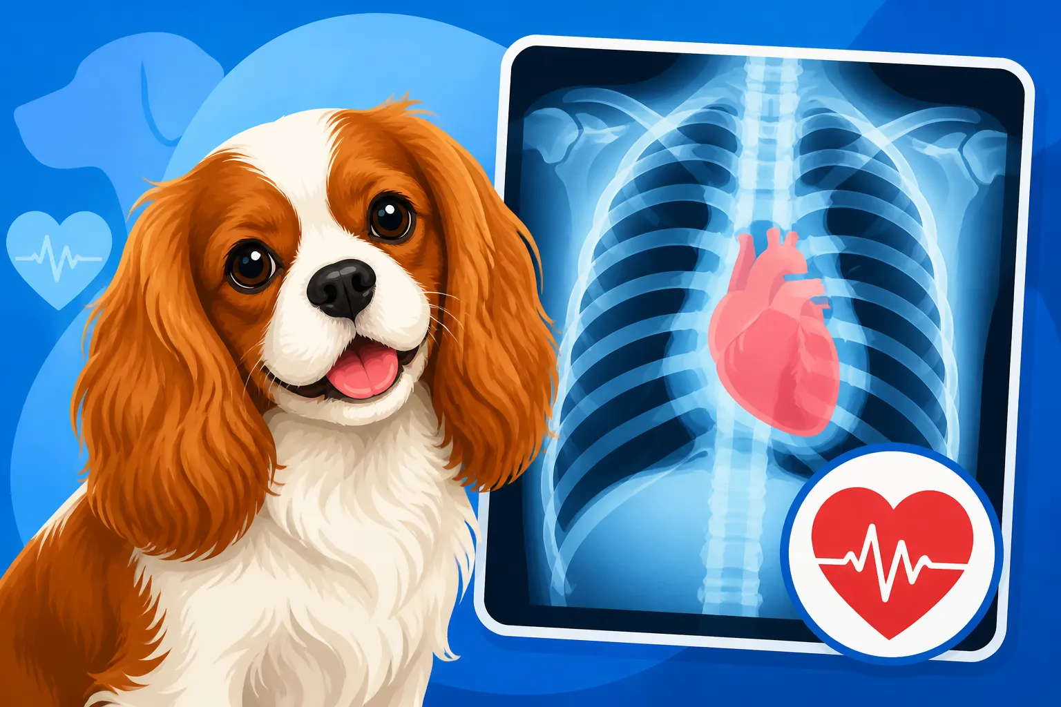
Chest X-Rays in a Cavalier Heart Workup
It's a heartbreaking moment when your beloved Cavalier King Charles Spaniel starts showing signs that might point to heart trouble. You notice a cough, maybe they’re tiring more easily, or perhaps your vet heard a new murmur during a routine check-up. The worry can be overwhelming. When your vet recommends a chest X-ray as part of a comprehensive heart workup, it can feel like a big, scary step. This guide is here to walk you through exactly why chest X-rays are so important for your Cavalier's heart health, what they show, and how the process usually goes, helping you understand this critical diagnostic tool.
Why Chest X-Rays are Crucial for Cavalier Heart Health
When your vet listens to your Cavalier's chest with a stethoscope, they might pick up a heart murmur – a whooshing sound that indicates turbulent blood flow. This often signals that something is amiss with the heart valves, most commonly the mitral valve in Cavaliers, leading to what we know as Mitral Valve Disease (MVD). But a stethoscope, while incredibly useful, can only tell part of the story. It tells your vet if there's a murmur, and sometimes even What a Heart Murmur Grade Means in a Cavalier, but it can't show the physical changes happening inside the chest. That's where chest X-rays become an indispensable tool. They provide a visual snapshot, allowing your veterinary team to see the actual size and shape of the heart and, crucially, to assess the lungs for any signs of fluid accumulation, a grave indicator of heart failure.
Visualizing the Heart and Lungs
A chest X-ray gives your vet a clear image of your Cavalier's thoracic cavity – that's the fancy name for the chest area where the heart and lungs reside. For a breed so predisposed to MVD, like our Cavaliers, this visual is paramount. The X-rays allow us to see if the heart is indeed enlarged, something that commonly occurs as MVD progresses. As the mitral valve weakens, the heart has to work harder, and the chambers can dilate and remodel, often getting bigger. This enlargement can sometimes put pressure on other structures in the chest, including the airways, leading to a cough that an owner might initially mistake for something else entirely.
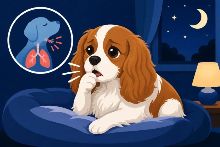
Beyond the heart itself, the X-ray is invaluable for scrutinizing the lungs. One of the most serious consequences of advanced MVD is the development of congestive heart failure (CHF), where fluid backs up into the lungs. This fluid, called pulmonary edema, severely impairs your dog's ability to breathe and is often what causes that distinctive cough associated with heart disease, especially Coughing at Night in a Cavalier. Seeing this fluid on an X-ray is a definitive sign of heart failure and requires immediate medical attention. It's the difference between hearing a problem and actually seeing its physical manifestation, guiding crucial treatment decisions.
Assessing Heart Enlargement (Cardiomegaly)
One of the primary reasons for taking chest X-rays in a Cavalier with a suspected heart condition is to objectively assess heart size. While your vet might have a good idea of heart enlargement just by listening and feeling, an X-ray provides a measurable, objective assessment known as the Vertebral Heart Score (VHS). This measurement involves comparing the size of the heart to the length of vertebrae in your dog's spine. It's a standardized method that allows veterinarians to quantify heart size and track changes over time.
A normal VHS for a Cavalier typically falls within a certain range. If your Cavalier's VHS is elevated, it indicates cardiomegaly, or an enlarged heart. This enlargement is a critical factor in determining the stage of your Cavalier's heart disease. For example, a Cavalier with a murmur but no significant heart enlargement on an X-ray might be categorized as ACVIM Stage B1, meaning they have MVD but no clinical signs or significant structural changes yet. However, if the X-ray reveals definite heart enlargement, even without fluid in the lungs, it moves them into Stage B2, a stage where starting specific heart medications like Pimobendan can significantly delay the onset of congestive heart failure. Knowing this precise measurement from the X-ray helps vets determine when to start Starting Heart Medication Before a Cavalier Coughs and manage the disease proactively.
What Your Vet Looks For on a Cavalier Chest X-Ray
Understanding what a veterinary cardiologist or your general practitioner is looking for on those black-and-white images can demystify the process and help you better grasp the conversation about your Cavalier's health. It’s not just about the heart; it’s about the whole picture within the chest cavity, as everything is interconnected.
Signs of Congestive Heart Failure (CHF)
The most urgent finding on a chest X-ray in a Cavalier with heart disease is the presence of pulmonary edema, which, as we mentioned, is fluid in the lungs – the hallmark of congestive heart failure. On an X-ray, this fluid typically appears as a hazy, fluffy, or patchy increase in density, particularly in certain areas of the lung fields. For left-sided heart failure, which is almost always the case with MVD, this fluid tends to accumulate in the perihilar region (around the main airways and blood vessels near the heart) and the caudal lung lobes (the rear portions of the lungs).
When your vet points this out on the X-ray, it means your Cavalier is struggling to breathe because their lungs are literally filling with fluid. This is a medical emergency requiring immediate treatment with diuretics to help remove the excess fluid and other medications to support heart function. Without an X-ray, it can sometimes be difficult to differentiate a heart-related cough from other types of coughs, like those caused by kennel cough or allergies. The X-ray offers a definitive answer, allowing for precise and life-saving intervention. It's a critical tool in understanding How Cavalier Heart Disease Progresses Stage by Stage.
Tracheal Compression and Bronchial Disease
Cavaliers, by their very nature, have relatively small airways, and when their heart significantly enlarges due to MVD, it can physically push against and compress the trachea (windpipe). This "tracheal compression" is another significant finding on a chest X-ray. You might see the trachea, which normally appears as a dark, air-filled tube, pushed upwards or flattened, especially where it passes over the enlarged heart. This compression can contribute significantly to your Cavalier's coughing, making it harder for them to clear their airways and breathe comfortably.
In addition to tracheal compression, X-rays can also reveal signs of chronic bronchial disease, which can sometimes coexist with heart issues or even mimic heart disease symptoms. This might appear as thickened bronchial walls, sometimes described as "doughnuts" or "tram lines" on the X-ray. While these findings might point to issues like chronic bronchitis, the X-ray helps your vet determine if the primary problem is cardiac, respiratory, or a combination of both. Differentiating between these causes is essential for effective treatment, ensuring your Cavalier gets the right medication for the right problem.
The Process of Getting a Cavalier Chest X-Ray
Understanding the practical side of getting a chest X-ray for your Cavalier can help ease some of the apprehension you might feel. It’s a common, relatively quick procedure, and your veterinary team will take every precaution to ensure your dog is comfortable and safe throughout.
Preparation and Positioning
When your vet recommends chest X-rays, they will usually ask you to drop off your Cavalier for a short period, typically an hour or two. This allows the veterinary technicians ample time to get the necessary images without you being present, which helps keep your dog calmer. Your Cavalier will be positioned carefully on an X-ray table, usually on their side (lateral views) and on their back or belly (ventrodorsal or dorsoventral views). These different angles are essential to get a comprehensive view of the heart and lungs, as certain structures can overlap in single projections.
Because it’s vital for your Cavalier to remain perfectly still for clear images, some dogs, especially those who are anxious or in pain, might require mild sedation. Your vet will discuss this with you beforehand. They use minimal sedation, just enough to relax your dog and ensure they cooperate, making the experience less stressful for everyone. The veterinary staff always wears protective gear, like lead aprons and thyroid shields, when taking X-rays, prioritizing their safety as well as your pet's. Knowing that dedicated professionals like those supported by pet insurance providers such as Healthy Paws are there to help can offer real peace of mind during potentially stressful times.
Understanding the Results and Next Steps
Once the X-rays are taken, your vet will carefully review the images, often with a cardiologist if the case is complex. They'll assess the heart size, look for pulmonary edema, and check for any other abnormalities. However, it's important to remember that X-rays are just one piece of the diagnostic puzzle. While they are excellent for showing heart size and fluid in the lungs, they don't provide details about the heart's internal structure or function. For that, your vet will likely recommend What a Cavalier Echocardiogram Shows. An echocardiogram is an ultrasound of the heart that allows the vet to see the valves, heart muscle, and blood flow in real-time.
The X-ray results, combined with findings from an echocardiogram, a physical exam, and possibly blood tests like A Cavalier Heart Blood Test Called NT-proBNP, help your vet accurately diagnose and stage your Cavalier's heart disease. This comprehensive approach ensures that the most appropriate treatment plan is put in place, whether that involves starting new medications, adjusting existing ones, or simply monitoring the condition. Your vet will explain all the findings to you, often showing you the images and helping you understand what they mean for your beloved Cavalier.
Integrating X-Ray Findings with Overall Heart Care and Costs
Living with a Cavalier King Charles Spaniel often means being prepared for the likelihood of heart disease. Integrating chest X-ray findings into the broader context of their care and understanding the associated costs is essential for responsible pet ownership.
The Role of X-Rays in Cavalier MVD Staging
The diagnostic journey for MVD in Cavaliers often follows the guidelines set by the American College of Veterinary Internal Medicine (ACVIM), which categorizes heart disease into specific stages (A, B1, B2, C, D). Chest X-rays play a particularly crucial role in differentiating between these stages, especially when determining if a Cavalier with a heart murmur has progressed to significant heart enlargement (Stage B2) or if they are in early congestive heart failure (Stage C).
Here’s a simplified look at how X-ray findings contribute to MVD staging:
| ACVIM Heart Disease Stage | Description | Key X-Ray Findings |
|---|---|---|
| Stage A | At risk, but no structural heart disease. | Normal heart size, no pulmonary edema. |
| Stage B1 | Murmur present, no radiographic or echocardiographic evidence of heart enlargement. | Normal heart size (normal VHS), no pulmonary edema. |
| Stage B2 | Murmur present, radiographic or echocardiographic evidence of significant heart enlargement. | Enlarged heart (elevated VHS), no pulmonary edema. Crucial for starting medication. |
| Stage C | Current or previous clinical signs of heart failure (e.g., coughing, difficulty breathing). | Enlarged heart, presence of pulmonary edema (fluid in lungs). Requires immediate treatment. |
| Stage D | End-stage heart failure, refractory to standard treatment. | Severely enlarged heart, persistent or recurrent pulmonary edema despite aggressive treatment. |
As you can see, the presence or absence of heart enlargement and pulmonary edema on an X-ray is often the defining factor for moving between stages B1, B2, and C. This staging directly impacts when your vet recommends starting certain medications, like Pimobendan for Stage B2, which can vastly improve your Cavalier's quality of life and potentially How Long a Cavalier Lives After Diagnosis.
Financial Considerations and Pet Insurance
It’s no secret that veterinary diagnostics and treatments can be expensive, and a full heart workup for a Cavalier is no exception. A chest X-ray, while essential, adds to the overall cost of care. You might be looking at fees for the X-rays themselves, the vet's interpretation, and potentially sedation. These costs quickly escalate when combined with consultations, blood tests, and an echocardiogram. Many Cavalier owners find themselves facing significant bills, especially once a heart murmur is detected and more intensive monitoring or medication becomes necessary. This is precisely why planning ahead with pet insurance can be a lifesaver.
Companies like Healthy Paws can provide invaluable support, helping to cover the costs of these diagnostic procedures and ongoing treatments. The key is to enroll your Cavalier in pet insurance Insuring a Cavalier Before the Murmur Arrives, ideally when they are a puppy and before any heart murmur is detected. Once a murmur appears, it's often considered a pre-existing condition, and many insurance policies won't cover treatment for it. Being proactive can save you immense financial stress down the line, allowing you to focus on your Cavalier's health without constantly worrying about What Cavalier Heart Disease Costs to Treat. Remember, annual check-ups are vital for early detection, and knowing what to expect from diagnostic tests like X-rays empowers you to make the best decisions for your beloved companion.
Disclaimer
The information provided in this guide is for informational purposes only and does not constitute veterinary medical advice. Always consult with a qualified veterinarian for any health concerns or before making any decisions about your pet's medical care.