What a Cavalier Echocardiogram Shows
It’s a moment no Cavalier owner wants to hear: "Your dog has a heart murmur." Your mind races, questions flood in, and a knot forms in your stomach. While a veterinarian can tell you they hear a murmur with a stethoscope, that sound alone can't explain what's truly going on inside your precious Cavalier's heart. This guide will walk you through what a Cavalier echocardiogram shows, why it's the most crucial diagnostic tool, and what those results mean for your beloved companion.
Understanding the Baseline: Why an Echocardiogram Matters
Hearing a heart murmur is often the first sign that something might be amiss with your Cavalier's heart. While a murmur definitely warrants attention, it's just the tip of the iceberg. Think of it like a car warning light — it tells you there’s a problem, but not what the problem is, or how severe. For a breed like the Cavalier King Charles Spaniel, prone to Mitral Valve Disease in the Cavalier King Charles Spaniel, understanding the underlying cause is paramount.
Beyond the Stethoscope: The Limitations of a Murmur Grade
When your vet listens to your Cavalier’s chest, they’re listening for unusual sounds. A murmur is literally the sound of turbulent blood flow, which can happen for various reasons. They might even give you a grade, from I (very faint) to VI (very loud and easily felt). You might hear something like, "Your Cavalier has a Grade II murmur," and while that gives you some information, it doesn't paint the full picture.
The frustration here is real. You've been told there's an issue, but you don't know the extent. Is it just a little leak? Is it serious? Will it get worse quickly? A murmur grade can sometimes correlate with the severity of the disease, but it's not a direct measure. A very loud murmur doesn't always mean severe heart disease, and a faint murmur doesn't necessarily mean it's benign. For instance, sometimes a very early, small leak can produce quite a loud sound. Conversely, a very sick heart in late-stage failure might actually have a quieter murmur because it’s struggling to pump enough blood to create turbulent flow. This is why solely relying on a stethoscope can lead to misinterpretations and unnecessary worry or, worse, a false sense of security. It leaves you feeling in the dark about what truly matters for your dog's health. You can read more about What a Heart Murmur Grade Means in a Cavalier.
The Ideal Scenario: Early Diagnosis for Better Outcomes
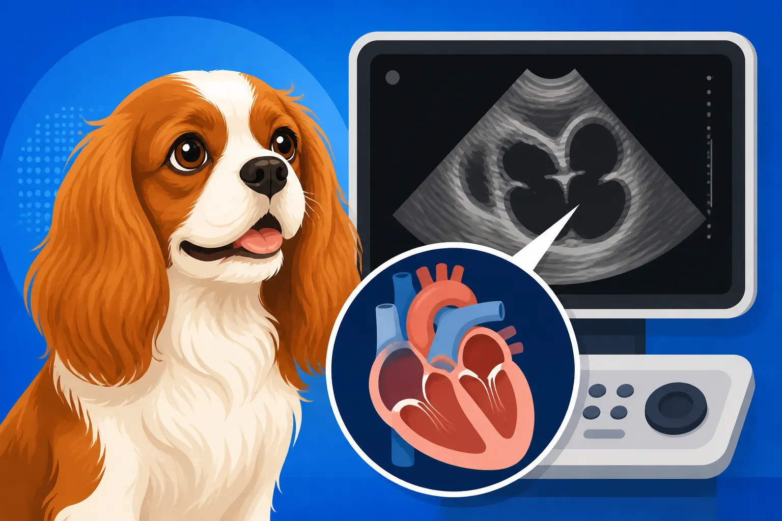
This is where the echocardiogram, often called an "echo," comes in. It’s a specialized ultrasound of the heart that allows a veterinary cardiologist to see the heart's structure, measure its chambers, assess valve function, and observe blood flow in real time. It's the definitive diagnostic tool that moves beyond mere sound. For Cavaliers, where heart disease is so prevalent—with Why Half of Cavaliers Have a Murmur by Five being a stark reality—early and accurate diagnosis is a game-changer.
Imagine knowing precisely what’s happening inside your Cavalier’s heart even before they show any symptoms. This isn't just about peace of mind; it's about proactively managing their health. Early diagnosis allows your vet and a cardiologist to establish a baseline, monitor progression, and, most importantly, intervene with medication at the optimal time. For example, specific medications like pimobendan have been proven to delay the onset of clinical signs of heart failure when started at the right stage. Waiting until your Cavalier is coughing or struggling to breathe often means the disease is already quite advanced. An echo helps avoid this delay, ensuring your beloved pet gets the best chance at a longer, more comfortable life. It means you can be ahead of the curve, not constantly playing catch-up, which is crucial when dealing with a progressive disease.
Key Structures and Measurements an Echocardiogram Reveals
An echocardiogram is incredibly detailed, offering a dynamic, real-time view of your Cavalier's heart. It's not just a snapshot; it's like a movie, showing the heart beating, valves opening and closing, and blood flowing. This allows the cardiologist to assess many critical aspects that a simple stethoscope check or even a chest X-ray simply cannot. This comprehensive view is why it's the cornerstone of diagnosing and managing heart disease in Cavaliers.
Visualizing the Mitral Valve: Where MVD Begins
For Cavaliers, the spotlight is almost always on the mitral valve. This tiny, two-leaflet valve separates the left atrium from the left ventricle, ensuring blood flows in one direction and doesn't back up. During an echocardiogram, the cardiologist meticulously examines these leaflets. They're looking for signs of thickening, irregularity, or prolapse – where the valve bulges backward into the left atrium during contraction.
Most importantly, they're looking for regurgitation, which is the hallmark of Mitral Valve Disease (MVD). This means the valve isn't closing tightly, allowing blood to leak backward into the left atrium. The echo can visually confirm this leak and, using Doppler technology, quantify its severity. They can see how much blood is backflowing, and how forcefully. This visual evidence of a leaky valve is what distinguishes a significant heart problem from an "innocent" murmur. Without this direct visualization, it's impossible to know Whether a Cavalier Murmur Always Means Disease. This detailed assessment provides the clear picture needed to understand the true impact of MVD on your dog's health, moving past the anxiety of 'what if' to the clarity of 'what is.'
Assessing Heart Chambers: Enlargement and Function
Beyond the valves, the echocardiogram provides precise measurements of the heart's chambers. The two most critical for MVD in Cavaliers are the left atrium (LA) and the left ventricle (LV). When the mitral valve leaks, blood backs up into the left atrium, causing it to enlarge. This enlargement is a critical indicator of disease progression. The cardiologist will measure the left atrium in relation to the aorta (LA:Ao ratio), which is a standard and highly important measurement. An enlarged left atrium signifies that the heart is working harder to compensate for the leaking valve.
The left ventricle, the main pumping chamber, is also closely scrutinized. Its size, wall thickness, and overall contractility (how well it squeezes) are all measured. The echo assesses how efficiently the heart is pumping blood to the body. Key metrics like Ejection Fraction (EF) and Fractional Shortening (FS) indicate the heart's pumping strength. If the heart muscle is weakening, these numbers will decline. These measurements are crucial for staging the disease and guiding treatment. If the left atrium or ventricle are significantly enlarged, it might be time to consider Pimobendan for a Cavalier With an Enlarged Heart. The comprehensive data gathered on chamber size and function offers a quantitative way to track the disease, moving from subjective observations to objective, measurable changes.
Measuring Blood Flow: Doppler Studies
One of the most powerful aspects of an echocardiogram is its ability to perform Doppler studies. This technology uses sound waves to measure the speed and direction of blood flow within the heart. It's how cardiologists visually confirm and quantify regurgitation. When the mitral valve leaks, the Doppler will show turbulent blood flowing backward into the left atrium. The color and pattern of this flow help determine the severity of the leak—a wider, more turbulent jet indicates more significant regurgitation.
Doppler also allows for the calculation of pressures within the heart chambers. For example, it can estimate the pulmonary artery pressure, which can become elevated in advanced heart disease due to blood backing up into the lungs. This information is vital for assessing the overall impact of MVD on the cardiovascular system and for identifying potential complications like pulmonary hypertension. It essentially turns the heart's complex fluid dynamics into a visible, measurable event, offering invaluable insights into its efficiency and any areas of distress. This detailed analysis helps tailor the treatment plan, ensuring that if Medication for a Cavalier With Heart Disease is needed, it’s the right type and dosage.
Here’s a summary of what an echocardiogram reveals:
| Heart Structure/Function | What the Echocardiogram Shows | Significance for Cavaliers |
|---|---|---|
| Mitral Valve | Thickening, prolapse of leaflets, regurgitation (leakage) | Direct evidence of MVD, severity of valve degeneration. |
| Left Atrium (LA) | Size (LA:Ao ratio), presence of enlargement | Primary indicator of MVD progression; enlargement suggests significant blood backup. |
| Left Ventricle (LV) | Size, wall thickness, pumping strength (Ejection Fraction, Fractional Shortening) | Assesses heart muscle function and overall efficiency of blood pumping. |
| Blood Flow (Doppler) | Direction and velocity of blood, turbulence, regurgitant jet | Confirms and quantifies leaks, estimates pressures within heart chambers. |
| Other Chambers/Valves | Integrity of other valves (aortic, tricuspid), right heart size | Rules out other heart conditions and assesses overall cardiac health. |
| Pericardium | Presence of fluid around the heart | Detects conditions like pericardial effusion, which can impair heart function. |
Interpreting the Results: What Your Cavalier's Echo Means
Getting the echocardiogram results can be a nerve-wracking experience, but understanding them is key to making informed decisions for your Cavalier's future. The cardiologist will translate all those complex images and measurements into a clear diagnosis and, importantly, a treatment plan. This report is far more comprehensive than a simple murmur grade and sets the stage for how you and your vet will manage your Cavalier's heart health moving forward.
Staging Mitral Valve Disease: ACVIM Guidelines
One of the most crucial outcomes of an echocardiogram is the accurate staging of Mitral Valve Disease, typically following the guidelines set by the American College of Veterinary Internal Medicine (ACVIM). These stages help standardize diagnosis and treatment protocols.
- Stage A: This is for Cavaliers (and other breeds) at high risk for heart disease but showing no structural changes or symptoms. Essentially, genetically predisposed, but currently healthy.
- Stage B1: A murmur is present, and the echo shows structural heart changes (like mitral valve thickening and regurgitation), but the heart itself is not significantly enlarged. This stage often doesn't require medication, but regular monitoring is crucial.
- Stage B2: This is a critical stage. A murmur is present, there are structural changes, and the heart, specifically the left atrium and left ventricle, are significantly enlarged due to the leak. Critically, there are still no outward symptoms like coughing or exercise intolerance. It is at this stage that evidence-based medicine strongly supports starting medication like pimobendan to delay the onset of clinical signs of heart failure. This is often the point when owners realize the disease is progressing, even without their dog acting sick.
- Stage C: Your Cavalier has heart disease and is showing clinical signs of heart failure, such as coughing, difficulty breathing, or exercise intolerance, and these signs are due to fluid accumulation or poor heart function. Medication combinations (like diuretics, pimobendan, ACE inhibitors) are essential for managing symptoms and improving quality of life.
- Stage D: This is end-stage heart failure, where your Cavalier's condition is refractory to standard treatments. It means the usual medications are no longer adequately controlling the symptoms. This stage requires intensive, often multimodal, medical management to try and maintain comfort.
Understanding these stages empowers you to grasp the seriousness of the diagnosis and the rationale behind the recommended treatments. It moves beyond a vague "heart problem" to a specific, actionable plan. These stages directly influence decisions about Starting Heart Medication Before a Cavalier Coughs and managing your dog's overall health.
Prognosis and Management: What Comes Next
The echocardiogram results, combined with the ACVIM staging, form the foundation of your Cavalier's long-term management plan. This plan isn't a one-and-done solution; MVD is a progressive disease. Your cardiologist will discuss the likely prognosis for your dog's specific stage, offering an estimate of How Long a Cavalier Lives After Diagnosis, while emphasizing that every dog is an individual.
Management typically involves:
- Medication: If your Cavalier is in Stage B2 or higher, medication will be prescribed. This might include pimobendan, diuretics, ACE inhibitors, or other drugs depending on the specific findings and stage. Adhering to the medication schedule is paramount.
- Monitoring: Regular re-evaluations, including follow-up echocardiograms and chest X-rays, will be crucial. For a Cavalier in Stage B2, an Annual Cardiologist Exams for a Cavalier might be recommended to track disease progression and adjust medication if needed. Owners are often taught how to count their dog's resting respiratory rate at home, a vital indicator of impending fluid in the lungs.
- Lifestyle Adjustments: This could mean dietary changes (e.g., low-sodium food if in later stages of heart failure), controlled exercise, and careful monitoring for any changes in behavior or breathing.
- Quality of Life: The ultimate goal is always to maintain the best possible quality of life for your Cavalier. This involves managing symptoms, minimizing distress, and keeping them comfortable and happy for as long as possible.
The costs associated with managing a Cavalier with heart disease, from the initial echo to ongoing medications and follow-up visits, can be substantial. This is where pet insurance can be a real lifesaver. Companies like Healthy Paws can help alleviate the financial burden of these essential diagnostics and treatments, allowing you to focus on your Cavalier's care without constant worry about the bills. It's often recommended to get insurance, like Healthy Paws, early on, ideally before any murmurs are detected, to ensure coverage for future conditions. This proactive step can make a huge difference in your ability to afford the best care when it's most needed. It’s also worth noting that the expenses can escalate significantly once The Bills That Land Once a Cavalier's Heart Fails become a reality.
Preparing for Your Cavalier's Echocardiogram
The thought of your dog undergoing any medical procedure can be daunting, but an echocardiogram is generally a very safe and non-invasive process. Knowing what to expect can help ease your mind and ensure a smooth experience for both you and your Cavalier. It’s a specialized test, so it's not something typically done at every veterinary clinic.
What to Expect During the Procedure
First and foremost, an echocardiogram is painless. Your Cavalier will lie on a special table with a cut-out section or be gently cradled by a technician. This allows the cardiologist to place a transducer (a small, handheld probe) directly against your Cavalier's chest, usually on the right side, where a small patch of hair might be shaved to ensure good contact and clear images. A special gel is applied to help the ultrasound waves penetrate effectively.
The procedure typically takes anywhere from 30 to 60 minutes. Most Cavaliers tolerate the process very well, especially with gentle handling, a quiet environment, and maybe a few treats. They might even fall asleep! Unlike some diagnostic procedures, sedation is often not required for an echocardiogram, unless a dog is particularly anxious or uncooperative. The cardiologist needs them to be still, but awake enough that their heart rate and breathing are natural, not suppressed by sedatives. You might be allowed to be present during the procedure to help keep your Cavalier calm, which can be a huge comfort to both of you. The goal is a stress-free experience, allowing for the clearest and most accurate imaging possible.
Finding a Board-Certified Veterinary Cardiologist
While your primary care veterinarian is excellent for general health, interpreting an echocardiogram and accurately diagnosing complex heart conditions like MVD requires specialized expertise. That's why it's highly recommended to seek out a board-certified veterinary cardiologist. These specialists have undergone extensive additional training, residencies, and rigorous examinations to become experts in the heart. They have the advanced equipment, training, and experience to perform and interpret the echo with the highest degree of accuracy.
You can typically find a board-certified cardiologist through referrals from your general veterinarian or by searching online directories provided by organizations like the American College of Veterinary Internal Medicine (ACVIM). Simply visit their website and use their 'Find a Specialist' tool. While a specialist visit might cost more upfront than a general vet visit, the precision of their diagnosis and tailored treatment plan can save money and heartache in the long run by avoiding misdiagnoses or inappropriate treatments. This is another area where having pet insurance, such as Healthy Paws, can be incredibly beneficial. Their coverage often includes specialist visits and advanced diagnostics, easing the financial burden and ensuring your Cavalier receives top-tier care from the most qualified professionals. When considering a puppy, remember that The Heart Testing a Cavalier Breeder Must Do involves these very specialists, showcasing their critical role from the very start.
Disclaimer
This article is intended for informational purposes only and does not constitute medical advice. Always consult with a qualified veterinary professional for any health concerns or before making any decisions related to your pet's care.